 Russian (Russia)
  English (United Kingdom)
 Статьи:
1. Volkov I.L., Serdobintsev P.Yu., Kononov, A.I. DNA-Stabilized Silver Nanoclusters with High Yield of Dark State, J. Phys. Chem. C, 2013, 117 (45), pp 24079–24308.
(PDF) Fluorophores with long-lived dark states have gained special attention in bioimaging microscopy. Such states in DNA-stabilized fluorescent clusters, a new class of bioprobes, have already been employed in optically modulated imaging scheme, though the formation efficiency of the dark state is estimated to be less than 1%. We measured quantum yield of the dark state for the calf thymus DNA-stabilized Ag nanocluster using nonlinear fluorescence saturation spectroscopy. The obtained value of the yield of 25% suggested very efficient formation of the nonemitting long-lived transient species of the studied silver cluster–DNA complex presumably of charge-transfer nature. This result seems to be promising in further creating polymer-stabilized Ag nanoclusters with specially designed structure providing high efficiency of the dark state formation.
2. Volkov, I.L.; Ramazanov, R.R.; Ubyvovk, E.V.; Rolich, V.I.; Kononov, A.I.; Kasyanenko, N.A. Fluorescent silver nanoclusters in condenced DNA, ChemPhysChem , 2013, 14 (15), 3543–3550.
(PDF) We study the formation and fluorescent properties of silver nanoclusters encapsulated in condensed DNA nanoparticles. Fluorescent globular DNA nanoparticles are formed using a dsDNA–cluster complex and polyallylamine as condensing agents. The fluorescence emission spectrum of single DNA nanoparticles is obtained using tip-enhanced fluorescence microscopy. Fluorescent clusters in condensed DNA nanoparticles appear to be more protected against destructive damage in solution compared to clusters synthesized on a linear polymer chain. The fluorescent clusters on both dsDNA and ssDNA exhibit the same emission bands (at 590 and 680 nm) and the same formation efficiency, which suggests the same binding sites. By using density functional theory, we show that the clusters may bind to the Watson–Crick guanine–cytosine base pairs and to single DNA bases with about the same affinity.
3. R.R. Ramazanov, and A.I. Kononov. Excitation Spectra Argue for Threadlike Shape of DNA-Stabilized Silver Fluorescent Clusters, J. Phys. Chem. C, 117 (36), 18681–18687.
|
(PDF) We calculate geometry and electronic excitation spectra of silver clusters containing 1–6 atoms bound to single DNA bases and also to dC3 cytosine oligomer using density functional theory method. We show that planar shaped silver clusters complexed with the bases exhibit nearly forbidden the lowest transitions and weak fluorescence ability. Their calculated spectra do not appear to match experimentally observed fluorescence excitation spectra of silver clusters on DNA and other polymers. On the contrary, threadlike-shaped silver clusters reveal intense the lowest transition, energy of which depends on the chain bend. We show the equilibrium structures of Ag3+ cluster stabilized by oxygen atoms on phosphates and in the minor groove of dC3 oligomer. The calculated excitation spectrum of Ag3+ cluster in the minor groove appears to be close to the experimental spectrum of green emitting clusters on cytosine oligomers. |
 |
4. Ramazanov R.R., Kononov A.I., Excitation spectra of thread-like silver clusters, in: Computational and Theoretical Modeling of Biomolecular Interactions .Moscow-Izhevsk: Institute of Computer Science, 2013, pp. 57-59. ISBN 978-5-4344-0126-5.
(PDF) It appears that just by means of variation of the bend in the chain of atoms in silver clusters one can get different low-energy peaks which cover the entire visible region (~1.7-3 eV) of the excitation spectra
5. Кононов А.И., Сибилева М.А., Михайлова Н.А., Грищенко А.Е., Двойное лучепреломление в потоке и оптическая анизотропия молекул поли-N-винилпирролидона, Высокомолек. Соед. А, 2013. 55, 4, 379-383.
Исследованы концентрационные зависимости ДЛП в потоке и вязкости растворов поли-N-винилпирролидона в воде и в бензиловом спирте.
Конференции:
- A. I. Kononov, Fluorescent metal nanodots as a special class of nanoobjects, 3-d International School on Surface Science, 23-29 September, 2013, Khosta (Sochi) Russia (Invited Talk).
- Ramazanov R.R., Kononov A.I., Excitation spectra of thread-like silver clusters, International Workshop “Computational and Theoretical Modeling of Biomolecular Interactions”, June 3 – 8, 2013, Dubna, Russia.
Статьи:
1. Ramazanov R.R., Kononov A.I., Heterogeneity of Threadlike Shape of DNA-Stabilized Silver Fluorescent Clusters, Biophysical Journal, 2014, 106(2), 806a.
DNA is widely used as a stabilizing matrix in the synthesis of fluorescent silver nanoclusters (AgNCs) possessing good biocompatibility and high brightness. We studied a group of threadlike shaped clusters up to 8 atoms having an elongated shape, so that each atom is connected with only one or two neighbors. Such the choice have been proved by recent calculations of electronic excitation spectra of threadlike shaped silver clusters showing intense the lowest transition, energy of which depends strongly on the chain bend.
2. Volkov I. L., Serdobintsev P. Yu., Kononov A. I., DNA-Stabilized Silver Nanoclusters with High Yield of Dark State Probed by Fluorescence Saturation Spectroscopy, Biophysical Journal, 2014, 106(2), 216a - 217a.
Polymer-stabilized silver nanoclusters have gained much attention as a promising higly fluorescent biolabels. While a wide palette of DNA-stabilized nanoclusters has been created, a little attention has been paid to their excited state properties. Meantime, dark long-lived non-emitting states have gained increasing interest in ultrahigh resolution microscopy, transient state imaging, and optically modulated microscopy in the last years. The photoinduced dark states are used to be probed by pump-probe spectroscopy.
3. Volkov I. L., Karpenko V. V., Kononov A. I., Single DNA-Shelled Silver Nanoclusters Probed by Tip Enhanced Fluorescence Spectroscopy, Biophysical Journal, 2014, 106(2), 395a.
Significant advances in nanoimaging have been made by development of aperture-less tip enhanced fluorescent microscopy, in which so called near field effect enhances the absorption and emission rates and allows overcoming the diffraction limit. Tip Enhanced Fluorescent Spectroscopy (TEFS) involves a combination of classical scanning tunneling microscope (STM) with optical confocal microscope where incident light is focused on the tip of metal probe enhancing electromagnetic field nearby.
4. Mikhailova N. A., Kononov A. I., Orientational order in the films of poly-N-vinylpyrrolidone, Journal of Optical Technology, 2014, 81, 11.
(PDF) When a polarized ray is obliquely incident on a film, rotating the film around an axis perpendicular to its surface results in birefringence, which is studied in this paper. Surface birefringence in polyvinylpyrrolidone films was investigated as a function of the angle of incidence of light and the film thickness. The results made it possible to estimate the orientational-order parameter of segments of the polyvinylpyrrolidone macromolecules in the near-surface layers, as well as to compare the orientational order as a function of the length of a statistical segment of the chains of the polymer studied here with the orientational order of polystyrene molecules.
Конференции:
- Ramazanov R. R., Maksimov D. A., Kononov A. I. Electronic spectra of non-canonical nucleic acid dimers, X international conference "Modern problems of polymer science", IMC, St. Petersburg, 2014. [Program]
- Ramazanov R. R., Maksimov D. A., Kononov A. I. Curved Nucleic Acid Patterns as a Target for Solar Radiation, 16th International congress on photobiology, Cordova, Argentina, 2014. [Abstract]
- Volkov I., Ramazanov R., Kononov A., Fast Long-Range Energy Transfer and Low-Lying Excited States in DNA, 16th International Congress on Photobiology, Cordova, Argentina, 2014. [Abstract]
- Kononov A., Volkov I., Ramazanov R., Serdobintsev P., Karpenko V., Usachov D., Adamchuk V., Structural and Optical Properties of DNA-Based Silver Fluorescent Nanoclusters, XII International Conference on Nanostructured Materials (NANO 2014), MSU, Moscow, 2014. [Abstract&Program]
- Сыч Т. С., Волков И. Л., Кононов А. И. Определение фотофизических параметров кластеров серебра на цитозин-содержащем олигонуклеотиде,1-я междисциплинарная конференция «Современные решения для исследования природных, синтетических и биологических материалов» , СПбГУ, С.-Петербург, [Abstract]
- Ramazanov R.R., Kononov A.I., Heterogeneity of Threadlike Shape of DNA-Stabilized Silver Fluorescent Clusters, Biophysical Society 58th Annual Meeting, San Francisco, California, USA, February 15-19, 2014.[Abstract]
- Volkov I. L., Serdobintsev P. Yu., Kononov A. I., DNA-Stabilized Silver Nanoclusters with High Yield of Dark State Probed by Fluorescence Saturation Spectroscopy, Biophysical Society 58th Annual Meeting, San Francisco, California, USA, February 15-19, 2014.[Abstract]
- Volkov I. L., Karpenko V. V., Kononov A. I., Single DNA-Shelled Silver Nanoclusters Probed by Tip Enhanced Fluorescence Spectroscopy, Biophysical Society 58th Annual Meeting, San Francisco, California, USA, February 15-19, 2014. [Abstract]
Статьи:
1. Vdovichev A.A., Ramazanov R.R., Kononov A.I. Structural model of gold and silver fluorescent clusters. Computer Research and Modeling, 2015, vol. 7, no. 2, pp. 263-269
(PDF) Данная работа посвящена систематическому исследованию равновесных конфигураций кластеров золота и серебра размером от 2 до 9 атомов. Для найденных самых низких по энергии конфигураций проводится расчет электронных спектров возбуждения. Все расчеты проводятся в рамках теории функционала плотности (ТФП) с использованием гибридного функционала B3LYP и псевдопотенциала LANL2DZ. На основании анализа полученных электронных спектров возбуждения делается попытка подтвердить предположение, что небольшие кластеры серебра и золота, способные к люминесценции, могут образовываться только в присутствии стабилизирующей матрицы. Авторы показывают, что без стабилизации в растворе образуются «плоские» и «сферические» конфигурации, спектры которых не соответствуют экспериментальным спектрам возбуждения люминесценции для комплексов «ДНК-кластер».
2. Ruslan R. Ramazanov, Dmitriy A. Maksimov, and Alexei I. Kononov. Noncanonical Stacking Geometries of Nucleobases as a Preferred Target for Solar Radiation. J. Am. Chem. Soc. 2015, 137(36), pp 11656-11665
|
(PDF) Direct DNA absorption of UVB photons in a spectral range of 290–320 nm of terrestrial solar radiation is responsible for formation of cyclobutane pyrimidine dimers causing skin cancer. Formation of UVB-induced lesions is not random, and conformational features of their hot spots remain poorly understood. We calculated the electronic excitation spectra of thymine, cytosine, and adenine stacked dimers with ab initio methods in a wide range of conformations derived from PDB database and molecular dynamics trajectory of thymine-containing oligomer. The stacked dimers with reduced interbase distances in curved, hairpin-like, and highly distorted DNA and RNA structures exhibit excitonic transitions red-shifted up to 0.6 eV compared to the B-form of stacked bases, which makes them the preferred target for terrestrial solar radiation. These results might have important implications for predicting the hot spots of UVB-induced lesions in nucleic acids. |
 |
3. Kasyanenko N, Lysyakova L, Ramazanov R, Nesterenko A, Yaroshevich I, Titov E, Alexeev G, Lezov A, Unksov I. Conformational and phase transitions in DNA-photosensitive surfactant solutions: Experiment and modeling. Biopolymers. 2015, 103(2):109-22
(PDF) DNA binding to trans- and cis-isomers of azobenzene containing cationic surfactant in 5 mM NaCl solution was investigated by the methods of dynamic light scattering (DLS), low-gradient viscometry (LGV), atomic force microscopy (AFM), circular dichroism (CD), gel electrophoresis (GE), flow birefringence (FB), UV–Vis spectrophotometry. Light-responsive conformational transitions of DNA in complex with photosensitive surfactant, changes in DNA optical anisotropy and persistent length, phase transition of DNA into nanoparticles induced by high surfactant concentration, as well as transformation of surfactant conformation under its binding to macromolecule were studied. Computer simulations of micelles formation for cis- and trans-isomers of azobenzene containing surfactant, as well as DNA-surfactant interaction, were carried out. Phase diagram for DNA-surfactant solutions was designed. The possibility to reverse the DNA packaging induced by surfactant binding with the dilution and light irradiation was shown
Конференции:
- Kononov A. I., Excitonic Mechanism for Long-Range Energy Transfer in Ag Cluster-DNA Complexes, ESP 2015 Congress, Aveiro (Portugal), 31 August - 4 September, 2015, OC317.
- Maksimov D. A., Ramazanov R. R., Kononov A. I. Non-Canonical Stacking Geometries of Nucleobases as Potential Hot Spots for Sunlight-Induced DNA Lesions. ESP 2015 Congress, Aveiro (Portugal), 31 August - 4 September, 2015. OC241
- Maksimov D.A., Ramazanov R.R., Kononov A.I. Electronic spectra of π-stacked pyrimidine dimers: an ab initio study. The 22nd International Conference Mathematics. Computing. Education. January 26 – 31, 2015, Pushchino, Russia. [Abstract]
Статьи:
1. R. R. Ramazanov, A. I. Kononov, A. M. Nesterenko, J. R. Shakirova, I. O. Koshevoy, E. V. Grachova, and S. P. Tunik. Luminescence Switching of a Gold–Copper Supramolecular Complex: A Physical Insight.J. Phys. Chem. C 2016, 120, 25541−25547
(PDF) Stimuli-responsive metal complexes are extensively studied due to their promising properties for various practical applications. In many cases, stimuli-induced response is provoked by phase transitions, which completely modify electronic properties of chromophoric center to give substantial modification of the phase emissive properties. However, complexity of the systems hinders theoretical calculations, which could be used to interpret the emission response, even at qualitative level. In this study, we apply a joint experimental and theoretical approach to model the luminescence switching of a vapochromic Au−Cu organometallic supramolecular complex in solution, amorphous, and crystal state. We have investigated luminescence spectra and emission anisotropy of the complex in solution, polymer gel, amorphous, and crystal states. The excitation and emission energies of the complex in vacuum and crystal state have been calculated using QM and QM/MM approaches. The results show that (1) the red shift of emission of the complex in solution is caused by the conformational relaxation in the T1 state and (2) the blue shift of crystalline state emission originates from the interactions of the complex with the crystal environment. The model elaborated to describe the phenomena observed may provide a practical way to analyze stimuli-induced luminescence switching in analogous systems.
2. Ruslan R. Ramazanov, Tomash S. Sych, Zakhar V. Reveguk, Dmitriy A. Maksimov, Artem A. Vdovichev, Alexei I. Kononov. Ag–DNA Emitter: Metal Nanorod or Supramolecular Complex? Journal of Physical Chemistry Letters, 2016, 7, pp 3560–3566
(PDF) Ligand-stabilized luminescent metal clusters, in particular, DNA-based Ag clusters, are now employed in a host of applications such as sensing and bioimaging. Despite their utility, the nature of their excited states as well as detailed structures of the luminescent metal–ligand complexes remain poorly understood. We apply a new joint experimental and theoretical approach based on QM/MM-MD simulations of the fluorescence excitation spectra for three Ag clusters synthesized on a 12-mer DNA. Contrary to a previously proposed “rod-like” model, our results show that (1) three to four Ag atoms suffice to form a partially oxidized nanocluster emitting in visible range; (2) charge transfer from Ag cluster to DNA contributes to the excited states of the complexes; and (3) excitation spectra of the clusters are strongly affected by the bonding of Ag atoms to DNA bases. The presented approach can also provide a practical way to determine the structure and properties of other luminescent metal clusters.
3. T. Sych, A. Polyanichko, A. Kononov, Albumin-stabilized fluorescent silver nanodots, J. Mol. Str. 2016, DOI: 10.1016/j.molstruc.2016.09.088
(PDF) Ligand-stabilized Ag nanoclusters (NCs) possess many attractive features including high fluorescence quantum yield, large absorption cross-section, good photostability, large Stokes shift and two-photon absorption cross sections. While plenty of fluorescent clusters have been synthesized on various polymer templates, only a few studies have been reported on the fluorescent Ag clusters on peptides and proteins. We study silver NCs synthesized on different protein matrices, including bovine serum albumin, human serum albumin, egg albumin, equine serum albumin, and lysozyme. Our results show that red-emitting Ag NCs can effectively be stabilized by the disulfide bonds in proteins and that the looser structure of the denatured protein favors formation of the clusters.
4. A A Vdovichev, T S Sych, Z V Reveguk, A A Smirnova, D A Maksimov, R R Ramazanov and A I Kononov. Structure of fluorescent metal clusters on a DNA template. Journal of Physics Conference Series, 2016, 741(1), pp 12069
(PDF) Luminescent metal clusters are a subject of growing interest in recent years due to their bright emission from visible to near infrared range. Detailed structure of the fluorescent complexes of Ag and other metal clusters with ligands still remains a challenging task. In this joint experimental and theoretical study we synthesized Ag-DNA complexes on a DNA oligonucleotide emitting in violet- green spectral range. The structure of DNA template was determined by means of various spectral measurements (CD, MS, XPS). Comparison of the experimental fluorescent excitation spectra and calculated absorption spectra for different QM/MM optimized structures allowed us to determine the detailed structure of the green cluster containing three silver atoms in the stem of the DNA hairpin structure stabilized by cytosine-Ag+-cytosine bonds.
5. Z Reveguk, E Ikonnikov and A Kononov. Electronic excitation energy transport in a DNA-Ag cluster complex. Journal of Physics Conference Series, 2016, 741(1), pp 12051
(PDF) We study electronic excitation transfer in the complexes of Ag clusters with oligonucleotides. Steady-state fluorescence excitation spectra show that the excitation energy is transferred to Ag cluster from all the 30 nucleobases of a 15-mer DNA duplex. This is in contrast to DNA-dyes complexes, where the energy transfer to the dye occurs from neighboring DNA bases only. Fluorescence decay curve for the DNA duplex shows that the process of energy transfer occurs within <100 fs. The obtained results suggest coherent excitonic type of the transfer rather than trivial Forster mechanism.
6. I. Volkov, T. Sych, P. Serdobintsev, Z. Reveguk, A. Kononov, Fluorescence saturation spectroscopy in probing electronically excited states of silver nanoclusters, Journal of Luminescence 2016, 172, 175-179.
(PDF) DNA-based fluorescent Ag clusters attract much attention due to their high brightness and sensitivity to environment, which can be used in chemical sensing and biosensing applications. Low chemical yield of the fluorescent Ag clusters in solution hinders measuring of their absorption cross-section in the ground state and rate constants in the excited states. We applied fluorescence saturation spectroscopy for determining the photophysical constants of an oligonucleotide-stabilized red emitting Ag cluster. Power dependencies of the fluorescence response to the pulse excitation with different pulse duration allowed us to obtain the values of absorption cross-section, dark states formation and deactivation rates of the cluster. The Ag fluorescent cluster exhibited a relatively high (17%) efficiency of a long-lived dark state formation. This feature of the Ag cluster might be further interesting for possible photodynamic and microscopy applications.
Конференции:
- Ramazanov R., Maksimov D., Vdovichev A., Kononov A. Luminescent silver clusters stabilized by DNA oligomer (5’-CCTCCTTCCTCC-3’): investigations by use of QM/MM approach. XXIII International Conference Mathematics. Computing. Education. Dubna, Russia, 25–30 January 2016 (abstract)
- Ramazanov R.R., Nesterenko A.M., Kononov A.I. Luminescence of Aurum-Cuprum ligand stabilized clusters in a solution and in crystal state computed by QM and QMMM techniques. XXIII International Conference Mathematics. Computing. Education. Dubna, Russia, 25–30 January 2016 (abstract)
- Evgenii Ikonnikov, Zakhar Reveguk, Aleksey Kononov. Electronic excitation energy transport in a DNA-Ag cluster complex. International School and Conference "Saint-Petersburg OPEN 2016" Academic University of the Russian Academy of Sciences. March 28 - 30, 2016, at St. Petersburg, Russia (abstract)
- Artem Vdovichev, Tomash Sych, Zakhar Reveguk, Anastasiya Smirnova, Dmitriy Maksimov, Alexey Kononov, Ruslan Ramazanov, Zahar Reveguk. Structure of fluorescent metal clusters on a DNA template.International School and Conference "Saint-Petersburg OPEN 2016" Academic University of the Russian Academy of Sciences. March 28 - 30, 2016, at St. Petersburg, Russia (abstract)
- Tomash Sych, Alexander Polyanichko, Alexey Kononov. Albumin-stabilized fluorescent silver nanodots. 33rd European Congress on Molecular Spectroscopy, 30 July-4 August 2016, Szeged, Hungary. (abstract)
- Ramazanov, RR; Maksimov, DA; Kononov, AI, Potential Hot Spots for Solar-Induced DNA Lesions, ASP Conference 2016: 21-26 May 2016, Tampa, USA, Book of abstracts, 14.3
- Reveguk, ZV; Ikonnikov, EO; Kononov, AI, Electronic excitation energy transfer in DNA and DNA-Ag cluster complexes, ASP Conference 2016: 21-26 May 2016, Tampa, USA, Book of abstracts, P2.23
Статьи:
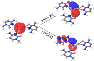
1. D.A. Maksimov, V.A. Pomogaev, A.I. Kononov, Excitation spectra of Ag3 –DNA bases complexes: a Benchmark Study, Chemical Physics Letters (2017), 673, pp 11-18. doi: http://dx.doi.org/10.1016/j.cplett.2017.01.074
|
(PDF) Assessment of different ab initio and TDDFT methods was studied for calculation of the excitation energies of the complexes of pyrimidine bases with positively charged Ag3+clusters. Performance of CIS, CIS(D), CC2, ADC(2), MP2, and TDDFT techniques with the use of different hybrid–GGA and meta–hybrid–GGA functionals and basis sets is studied. We found that M06–2X functional shows good accuracy in comparison with the ADC(2) ab initio method and that the geometry optimization approach can strongly affect the excitation spectra of the complexes. Our results may have important implications for further studies of ligand–stabilized silver nanoclusters. |
 |
2. Ivan L Volkov, Anastasiya Smirnova, Anna A Makarova, Zakhar V Reveguk, Ruslan R Ramazanov, Dmitry Yu Usachov, Vera K Adamchuk, Alexei I Kononov. DNA with Ionic, Atomic and Clustered Silver: An XPS Study. The Journal of Physical Chemistry B, 2017, DOI: 10.1021/acs.jpcb.6b11218
(PDF) The rapidly developing field of bionanotechnology requires detailed knowledge of the mechanisms of interaction between inorganic matter and biomolecules. Under conditions different from those in an aqueous solution, however, the chemistry of these systems is elusive and may differ dramatically from their interactions in vitro and in vivo. Here, we report for the first time a photoemission study of a metal silver–DNA interface, formed in vacuo, in comparison with DNA–Ag+ and fluorescent DNA–Ag complexes formed in solution. The high-resolution photoelectron spectra reveal that in vacuo silver atoms interact mainly with oxygen atoms of the phosphodiester bond and deoxyribose in DNA, in contrast to the behavior of silver ions, which interact preferentially with the nitrogen atoms of the bases. This offers new insight into the mechanism of DNA metallization, which is of importance in creating metal–bio interfaces for nanotechnology applications.
3. Ivan L. Volkov, Zakhar V. Reveguk, Pavel Yu. Serdobintsev, Ruslan R. Ramazanov and Alexei I. Kononov. DNA as UV lighy-harvesting antenna. Nucleic Acids Research, 2017 doi: 10.1093/nar/gkx1185
(PDF) The ordered structure of UV chromophores in DNA resembles photosynthetic light-harvesting complexes in which quantum coherence effects play a major role in highly efficient directional energy transfer. The possible role of coherent excitons in energy transport in DNA remains debated. Meanwhile, energy transport properties are greatly important for understanding the mechanisms of photochemical reactions in cellular DNA and for DNA-based artificial nanostructures. Here, we studied energy transfer in DNA complexes formed with silver nanoclusters and with intercalating dye (acridine orange). Steady-state fluorescence measurements with two DNA templates (15-mer DNA duplex and calf thymus DNA) showed that excitation energy can be transferred to the clusters from 21 and 28 nucleobases, respectively. This differed from the DNA–acridine orange complex for which energy transfer took place from four neighboring bases only. Fluorescence up-conversion measurements showed that the energy transfer took place within 100 fs. The efficient energy transport in the Ag–DNA complexes suggests an excitonic mechanism for the transfer, such that the excitation is delocalized over at least four and seven stacked bases, respectively, in one strand of the duplexes stabilizing the clusters. This result demonstrates that the exciton delocalization length in some DNA structures may not be limited to just two bases.
Статьи:
1. Z. Reveguk, R. Lysenko, R. Ramazanov, A. Kononov, Ultrafast fluorescence dynamics of DNA-based silver clusters, Phys. Chem. Chem. Phys., 2018, 20, 28205-28210, doi:10.1039/C8CP05727C
2. T. S. Sych, A. A. Buglak, Z. V. Reveguk, V. A. Pomogaev, R. R. Ramazanov, A. I. Kononov, Which Amino Acids are Capable of Nucleating Fluorescent Silver Clusters in Proteins? J. Phys. Chem. C, 2018, 122 (45), 26275–26280, doi:10.1021/acs.jpcc.8b08979
3. T. S. Sych, Z. V. Reveguk, V. A. Pomogaev, A. A. Buglak, A. A. Reveguk, R. R. Ramazanov, N. M. Romanov, E. V. Chikhirzhina, A. M. Polyanichko, A. I. Kononov, Fluorescent Silver Clusters on Protein Templates: Understanding Their Structure, J. Phys. Chem. C, 2018, 122 (51), 29549–29558, doi:10.1021/acs.jpcc.8b08306
4. A.A. Buglak, A.I. Kononov. Triplet state generation by furocoumarins revisited: a combined QSPR/DFT approach. New J. Chem., 2018, 42, 14424-14432, doi:10.1039/C8NJ03002B
Статьи:
1. A. A. Buglak, V. A. Pomogaev, A. I. Kononov. Calculation of absorption spectra of silver-thiolate complexes. Computer Research and Modeling, 2019 doi:10.1039/C8CP05727C
2. A. A. Buglak, R. R. Ramazanov, A. I. Kononov. Silver Cluster-Amino Acid Interactions: a Quantum-Chemical Study. Amino Acids, 2019, 51, 855–864, doi:10.1007/s00726-019-02728-z
3. A.A. Buglak, T.A. Telegina, E.A. Vorotelyak, A.I. Kononov. Theoretical study of photoreactions between oxidized pterins and molecular oxygen. J Photochem Photobiol, 2019, 372, 254-259, doi:10.1016/j.jphotochem.2018.12.002
4. Buglak, A.A., Pomogaev, V.A., Kononov, A.I., Predicting absorption spectra of silver-ligand complexes, Int J Quantum Chem. 2019; e25995, doi:10.1002/qua.25995
5. Z. V. Reveguk, E. V. Khoroshilov, A. V. Sharkov, V. A. Pomogaev, A. A. Buglak, A. N. Tarnovsky, A. I. Kononov, Exciton Absorption and Luminescence in i-Motif DNA, Sci. Rep. 2019; 9: 15988, doi:10.1038/s41598-019-52242-1
6. Sych T.S., Polyanichko A.M., Plotnikova L.V., Kononov A.I., Luminescent silver nanoclusters in probing immunoglobulins and serum albumins in protein mixtures, Anal. Methods, 2019, 11, 6153-6158, doi:10.1039/C9AY02054C
7. T. S. Sych, A. M. Polyanichko, R. R. Ramazanov, A. I. Kononov, tRNA as a stabilizing matrix for fluorescent silver clusters: photophysical properties and IR study, Nanoscale Adv., 2019,1, 3579-3583, doi:10.1039/C9NA00112C
Статьи:
1. Buglak, A.A., Kononov, A.I., Comparative study of gold and silver interactions with amino acids and nucleobases, RSC Advances, 2020, 10(56), 34149-34160, doi:10.1039/D0RA06486F
2. Pomogaev V.A., Kliuev P.N., Ramazanov R.R., Kononov A.I. Combined Quantum-Classical Simulation of Photoinduced Electronic Density Redistribution from Biopolymer Segments to Photochromic Probes //Russ. Phys. J. 2020. Vol. 63, № 8. P. 1386, doi:10.1007/s11182-020-02182-5
3. A V Mizincev, T S Sych, A M Polyanichko, L V Plotnikova, A D Garifullin, S V Voloshin, A I Kononov Fluorescent Ag-Nanoclusters for Evaluation of Serum Albumin and Immunoglobulin Content in Protein Mixtures J. Phys.: Conf. Ser. 2020 1695 012061, doi:10.1088/1742-6596/1695/1/012061
4. M A Kapitonova, Z V Reveguk and A I Kononov Revealing a possible sensor mechanism of DNA - based silver nanoclusters J. Phys.: Conf. Ser. 2020 1695 012058, doi:10.1088/1742-6596/1695/1/012058
5. Ya V Chuiko, V G Kubenko, Z V Reveguk, M A Kapitionova and A I Kononov Formation of luminescent nanoclusters by etching silver nanoparticles with biomolecules 2020 J. Phys.: Conf. Ser. 1695 012196, doi:10.1088/1742-6596/1695/1/012196
Статьи:
1. Reveguk Z.V., Pomogaev V.A., Kapitonova M.A., Buglak A.A., Kononov A.I. Structure and Formation of Luminescent Centers in Light-up Ag Cluster-Based DNA Probes //J. Phys. Chem. C. 2021, 125, 6, 3542–3552, doi:10.1021/acs.jpcc.0c09973
2. Buglak, Andrey A., et al. "Autoxidation and photooxidation of tetrahydrobiopterin: A theoretical study." Free Radical Research 55.5 (2021): 499-509, https://doi.org/10.1080/10715762.2020.1860213
3. Vechtomova, Yuliya L., et al. "UV Radiation in DNA Damage and Repair Involving DNA-Photolyases and Cryptochromes." Biomedicines 9.11 (2021): 1564, https://doi.org/10.3390/biomedicines9111564
4. Buglak, Andrey A., et al. "Quantitative Structure‐Property Relationship Modelling for the Prediction of Singlet Oxygen Generation by Heavy‐Atom‐Free BODIPY Photosensitizers." Chemistry–A European Journal 27.38 (2021): 9934-9947, https://doi.org/10.1002/chem.202100922
Статьи:
1. Buglak, Andrey A., and Alexei I. Kononov. "Silver Cluster Interactions with Tyrosine: Towards Amino Acid Detection." International journal of molecular sciences 23.2 (2022): 634, https://doi.org/10.3390/ijms23020634
2. Tatsiana Mikulchyk, Safakath Karuthedath, Catherine S.P. De Castro, Andrey A. Buglak, Aimee Sheehan, Aaron Wieder, Frédéric Laquai, Izabela Naydenova, Mikhail A. Filatov. Charge transfer mediated triplet excited state formation in donor–acceptor–donor BODIPY: Application for recording of holographic structures in photopolymerizable glass. Journal of Materials Chemistry C 10:32 (2022): 11588-11597, https://doi.org/10.1039/D2TC02263J
3. Т.А. Телегина, Ю.Л. Вечтомова, М.С. Крицкий, А.С. Низамутдинов, Э.И. Мадиров, Д.А. Макарова, А.А. Буглак. "Фотоокисление тетрагидробиоптерина как основа фототерапии витилиго". Оптика и спектроскопия, 130:5 (2022): 761, http://dx.doi.org/10.21883/OS.2022.05.52432.14-22
4. Andrey A. Buglak, Alexei I. Kononov. "Silver cluster interactions with Pterin: Complex structure, binding energies and spectroscopy". Spectrochimica Acta Part A: Molecular and Biomolecular Spectroscopy, 297 (2022): 121467, https://doi.org/10.1016/j.saa.2022.121467
5. Andrey A. Buglak, Marina A. Kapitonova, Yulia L. Vechtomova, Taisiya A. Telegina. "Insights into Molecular Structure of Pterins Suitable for Biomedical Applications". International Journal of Molecular Sciences, 23:23 (2022): 15222, https://doi.org/10.3390/ijms232315222
6. V.A. Pomogaev, H.J. Lee, E. Goh, O.N. Tchaikovskaya, A.I. Kononov, P.V. Avramov. "Electronically Excited States in Model Complexes of Noble Metal Clusters with Carbon Nanodots". Russian Physics Journal, 64 (2022): 2076-2081, https://doi.org/10.1007/s11182-022-02558-9
Статьи:
1. Platon P. Chebotaev, Vitaly Yu. Plavskii, Alexei I. Kononov, Andrey A. Buglak. "Pterin interactions with gold clusters: A theoretical study". Dyes and Pigments, 216 (2023): 111323, https://doi.org/10.1016/j.dyepig.2023.111323
2. Tomash S. Sych, Alexander M. Polyanichko, Andrey A. Buglak, Alexei I. Kononov. "Quantitative determination of albumin and immunoglobulin in human serum using gold nanoclusters". Spectrochimica Acta Part A: Molecular and Biomolecular Spectroscopy, 298 (2023): 122796, https://doi.org/10.1016/j.saa.2023.122796
3. Zakhar V. Reveguk, Tomash S. Sych, Alexander M. Polyanichko, Yana V. Chuiko, Andrey A. Buglak, Alexei I. Kononov. "Rapid and selective colorimetric determination of L-DOPA in human serum with silver nanoparticles". Spectrochimica Acta Part A: Molecular and Biomolecular Spectroscopy, 299 (2023): 122810, https://doi.org/10.1016/j.saa.2023.122810
Статьи:
1. Jael R. Neyra Recky, Maira Gaspar Tosato, Andrey A. Buglak, M. Laura Dántola, Carolina Lorente. "Photosensitized isomerization of resveratrol: Evaluation of energy and electron transfer pathways". Free Radical Biology and Medicine, 216 (2024): 50-59, https://doi.org/10.1016/j.freeradbiomed.2024.01.038
2. T.A. Telegina, Yu.L. Vechtomova, M.S. Kritsky, A.S. Nizamutdinov, E.I. Madirov, D.A. Makarova, A.A. Buglak "Photooxidation of Tetrahydrobiopterin as the Basis of Vitiligo Phototherapy". Optics and Spectroscopy, 131 (2023): 607-613, https://doi.org/10.1134/S0030400X2305017X
3. Andrey A. Buglak, Alexei I. Kononov "Interactions of deprotonated phenylalanine with gold Clusters: Theoretical study with prospects for amino acid detection". Spectrochimica Acta Part A: Molecular and Biomolecular Spectroscopy, 311 (2024): 124004, https://doi.org/10.1016/j.saa.2024.124004
4. Zakhar V. Reveguk, Evgeny V. Khoroshilov, Andrey V. Sharkov, Vladimir A. Pomogaev, Andrey A. Buglak and Alexei I. Kononov "Excited States in Single-Stranded and i-Motif DNA with Silver Ions". Journal of Physical Chemistry B, 128 (2024): 4377-4384, https://doi.org/10.1021/acs.jpcb.4c01127
5. Tomash S. Sych, Nikolai V. Shekhovtsov, Andrey A. Buglak and Alexei I. Kononov "Amino acid-stabilized luminescent gold clusters for sensing pterin and its analogues". Analytical Methods, 16 (2024): 4607-4618, https://doi.org/10.1039/D4AY00700J
6. Daria A. Belinskaia, Anastasia A. Batalova, Polina A. Voronina, Vladimir I. Shmurak, Mikhail. A. Vovk, Alexander M. Polyanichko, Tomash S. Sych, Kamila V. Samodurova, Vasilisa K. Antonova, Anastasia A. Volkova, Bogdan A. Gerda, Richard O. Jenkins, Nikolay V. Goncharov "Modulation of Albumin Esterase Activity by Warfarin and Diazepam". International Journal of Molecular Science, 25 (2024): 11543, https://doi.org/10.3390/ijms252111543
7. Andrey A. Buglak, Minh Tho Nguyen "Interactions of coinage metal nanoclusters with low-molecular-weight biocompounds". Biophysical Reviews, 16 (2024): 441-477, https://doi.org/10.1007/s12551-024-01200-x
8. Y.M. Hamdan, D.A. Makarova, N.I. Shamsutdinov, P.V. Zelenikhin, A.S. Nizamutdinov, A.A. Buglak, T.A. Telegina "Study of the Effect of UV Laser Pulse Duration and Wavelength on Fibroblasts". Physics of Particles and Nuclei Letters, 21 (2024): 835-838, https://doi.org/10.1134/S1547477124700717
9. Platon P. Chebotaev, Andrey A. Buglak, Aimee Sheehan, Mikhail A. Filatov "Predicting fluorescence to singlet oxygen generation quantum yield ratio for BODIPY dyes using QSPR and machine learning". Physical Chemistry Chemical Physics, 26 (2024): 25131-25142, https://doi.org/10.1039/D4CP02471K
Articles:
1. Varvara G. Kubenko, Vladimir A. Pomogaev, Andrey A. Buglak, Alexei I. Kononov. "Photophysics of 5,6,7,8-tetrahydrobiopterin on a femtosecond time-scale". Journal of Photochemistry and Photobiology B: Biology, 265 (2025): 113134, https://doi.org/10.1016/j.jphotobiol.2025.113134